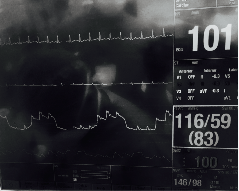
Example of Pulsus Paradoxus as seen with Pericardial Effusion/Tamponade
WakeMed Laser Lead Extraction Guidelines
Anesthetic Type:
· General Anesthetic ETT
Vascular Access:
· Peripheral IV (large bore preferred)
· Central venous lines (2 total)
o Right Internal Jugular Introducer placed by Anesthesiologist
o Femoral Vein 8.5Fr Cordis Line placed by Surgeon
§ Note: connect large bore sterile line to this
· Arterial line (with pulse oximeter on contralateral side)
Monitoring:
· IF LV ejection fraction less than 30% consider Cerebral oximetery if avail
· BIS Monitor
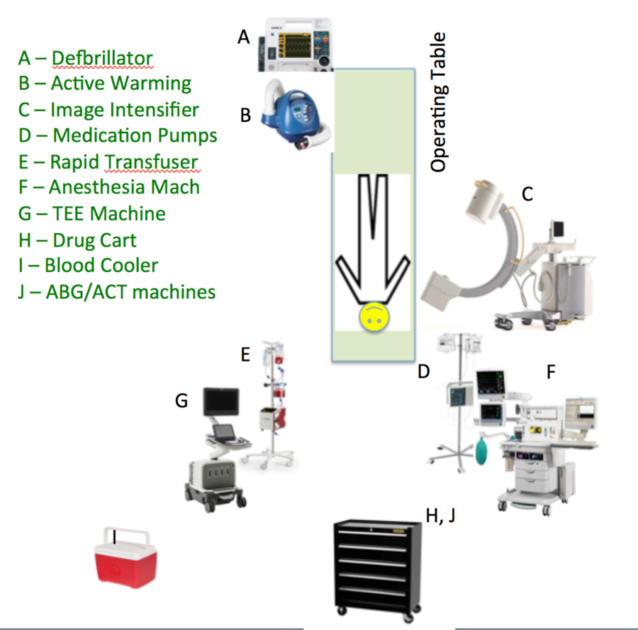
Special Equipment:
· TEE Machine (Does not need to be 3D machine)
· Hybrid room: obtain from Heart center. Ask cath lab staff.
· Main OR: use OR machine
· Defibrillator pads on sides of chest before induction.
· Test Sternotomy Saw, out loud, prior to incision
Blood Status:
· Blood should be present and checked in room prior to procedure start
· No TEG unless clinically indicated (rare)
· Call for Belmont rapid transfuser immediately if tamponade diagnosed. If lead is known to be particularly embedded, have Belmont connected to patient.
Phases of Surgery:
1) Initiation of anesthetic and establishment of vascular access
2) Echo to assess leads, cardiac status, presence of prior effusion
3) Surgeon will place Femoral Venous line and hand off end for connection.
4) Cardiologist/Surgeon will make incision at pacemaker pocket and thread laser catheter onto lead.
5) ***Page Attending Anesthesiologist prior to laser being used. Wear laser glasses and lead for fluoroscopy.
6) Cardiologist and surgeon will use tool to remove lead while Anesthesiologist looking at TEE. Expect 10 minutes per lead extracted.
7) Echo will be used to look for signs of tamponade, pericardial effusion or valve dysfunction throughout lead removal.
8) Note: Rising CVP and Pulsus Paradoxus on Arterial line are specific for diagnosis and may be seen before echo reveals findings.
9) New lead may or may not be placed after old one removed.
10) Incision is closed. Echo images obtained by anesthesiologist at intervals. TEE may be removed when deemed ok and stable by Anesthesiologist.
Pericardial effusion on TEE – Decompressed RV and LV
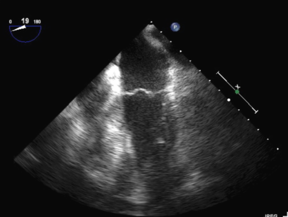
Four Chamber View on TEE of biventricular and biatrial volume reduction
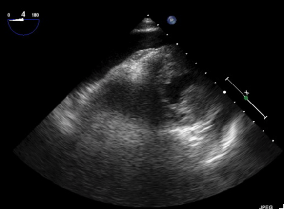
Transgastric View on TEE of pericardial effusion on posterior wall with resultant Chamber collapse